AbMole Mini-Lecture | Y-27632 – The Remarkable ROCK Inhibitor as a Key Bioactive Molecule in Primary Cell, Stem Cell, and Organoid Culture

Y-27632 is a highly selective, ATP-competitive inhibitor of Rho-associated coiled-coil containing protein kinase (ROCK), targeting both ROCK1 and ROCK2. It is an essential small-molecule compound used in primary cell, stem cell, and organoid culture.  

I. Molecular Mechanism of Action of Y-27632

Y-27632 reversibly inhibits ROCK kinase activity by competitively binding to its ATP-binding pocket. Within cells, ROCK functions downstream of the Rho GTPase signaling pathway and plays critical roles in regulating cellular contraction, migration, adhesion, and cell cycle progression. Upon activation of Rho GTPase, ROCK is activated and subsequently phosphorylates various substrates, including MYPT1, the regulatory subunit of myosin light chain phosphatase (MLCP). Phosphorylation of MYPT1 reduces MLCP activity, leading to increased phosphorylation of myosin light chain (MLC), which in turn promotes cytoskeletal contraction and remodeling. By inhibiting ROCK activity, Y-27632 prevents MYPT1 phosphorylation, maintains MLCP activity, and promotes MLC dephosphorylation, ultimately resulting in cytoskeletal relaxation and modulation of related cellular behaviors.  

II. Application of Y-27632 in Primary Cell Culture

In vivo, cells reside within a complex and intricate extracellular matrix (ECM) network. Through integrins, cells bind to specific ECM components (e.g., collagen, fibronectin, laminin), which not only provide physical anchorage but also continuously transmit critical survival signals. These signals activate pathways such as PI3K/Akt and FAK (focal adhesion kinase) to inhibit apoptotic programs. Upon cell dissociation, integrin-mediated signaling is disrupted, leading to Rho GTPase activation and subsequent robust activation of ROCK. Activated ROCK phosphorylates myosin light chain (MLC) and inhibits MLC phosphatase, resulting in sustained MLC phosphorylation. This induces a sharp increase in actomyosin contractility and triggers apoptosis, a process termed anoikis. As a potent ROCK inhibitor, Y-27632 significantly reduces MLC phosphorylation levels and restores normal myosin contractile activity upon ROCK inhibition. This helps maintain a relatively stable cytoskeletal structure even after detachment from the ECM, prevents apoptotic membrane blebbing, and facilitates cell reattachment.  

Case Study

ACS Biomater Sci Eng. 2025 Jan 13;11(1):451-462.

In this study, researchers from Erasmus Medical Center in Rotterdam, the Netherlands, designed a 3D-printed porous flow plate with high biocompatibility to support microfluidic cell culture. To validate the utility of this plate, primary human bronchial epithelial cells (hPBEC) and primary human pulmonary microvascular endothelial cells (hMVEC) were co-cultured on the apical and basolateral sides of the plate, respectively. The plate supported mucociliary differentiation of hPBEC and rapid proliferation of hMVEC. In both primary cell cultures, Y-27632 (provided by AbMole, M1817) was used at a concentration of 5 μM.  
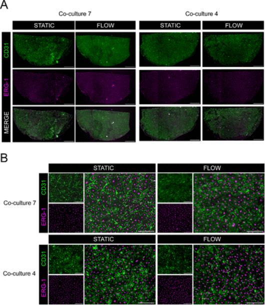
Figure 2. Co-culture of primary hMVEC and hPBEC.
AbMole Mini-Lecture | Vorinostat (SAHA, MK0683): Applications of a Histone Deacetylase (HDAC) Inhibitor in Epigenetics, Cancer, and Neurobiology

Vorinostat (SAHA, MK0683, AbMole, M1780) is a broad-spectrum, pan-histone deacetylase inhibitor (HDACi). Belonging to the succinylhydroxamic acid class of compounds, it directly binds to and inhibits HDAC activity, leading to the accumulation of acetylated histones, thereby regulating gene expression and affecting multiple signaling pathways. Additionally, Vorinostat (MK0683) exhibits bioactivities including cell cycle arrest, apoptosis, and inhibition of angiogenesis. Studies have shown that Vorinostat bidirectionally regulates stress response genes (e.g., sod-3, hsp-16.2, skn-1). It upregulates these genes at low concentrations but exhibits inhibitory or neutral effects at high concentrations (10 μM), demonstrating dose-dependent epigenetic regulatory properties [1]. Vorinostat also induces cell cycle arrest and programmed cell death by downregulating the Akt signaling pathway [2]. Furthermore, Vorinostat downregulates multiple epigenetic regulatory enzymes (e.g., EZH2, SUV39H1/2, DOT1L), indicating its broad impact on the epigenetic regulatory network [3].

In research applications, Vorinostat (SAHA) has shown multifaceted potential. First, it inhibits the proliferation of various tumor cells, including colon cancer cells (HCT116, HT29) [4], pancreatic cancer cells (AsPC-1) [5], hepatocellular carcinoma cells (LCL-PI 11) [5], and head and neck squamous cell carcinoma (HNSCC) cells [6]. Vorinostat is also used to inhibit parasites, with the mechanism similarly involving HDAC inhibition in protozoan cells [7]. Notably, the activity status of tissue transglutaminase 2 (TG2) affects cellular sensitivity to Vorinostat. Inhibition of TG2 activity enhances the anti-proliferative effect of Vorinostat, whereas TG2 overexpression confers resistance, identifying TG2 as a key target in Vorinostat tolerance mechanisms [8]. In the field of neuroprotection, Vorinostat reverses amyloid β protein -induced neural damage, an activity involving regulation of the AKT-MDM2-p53 pathway [9].

AbMole provides global researchers with high-purity, high-bioactivity inhibitors, cytokines, human monoclonal antibodies, natural products, fluorescent dyes, peptides, compound libraries, antibiotics, and other research reagents, widely cited in numerous publications and patents worldwide.

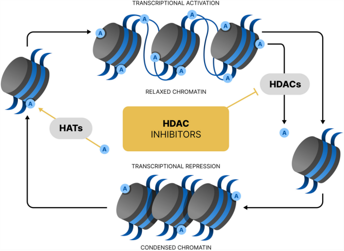
Transcriptional activity of histone deacetylase (HDAC) inhibitors[10].

Case Study

BMC Cancer. 2016 Nov 7;16(1):857.

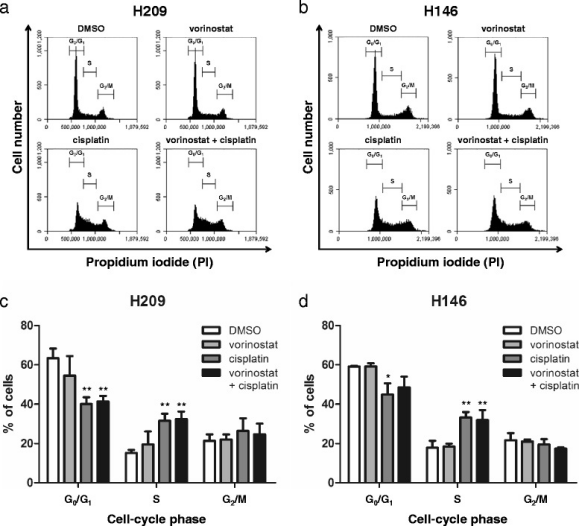
In this study, researchers investigated the inhibitory effect of Vorinostat on small cell lung cancer (SCLC) and the development of combination therapy strategies. Through in vitro (H209, H146 cell lines) and in vivo (H209 xenograft nude mouse model) experiments, the study demonstrated that Vorinostat (an HDAC inhibitor), when combined with Cisplatin or with Etoposide (VP-16-213), more significantly inhibited tumor cell viability, induced apoptosis (via caspase-3 activation and PARP cleavage), and induced S-phase cell cycle arrest compared to single-agent treatments. The combination also increased acetylation levels of histone H3 and α-tubulin while persistently inhibiting thymidylate synthase (TS) expression. The core research compound, Vorinostat (SAHA, MK0683, AbMole, M1780), was provided by AbMole. In 2014, two inhibitors from AbMole were used in in vivo studies by the Spanish National Center for Cardiovascular Research (CNIC) and Columbia University, leading to research publications in Nature and Nature Medicine.

 Cell cycle progression analysis of vorinostat in combination with cisplatin in SCLC cells[11].

References and Acknowledgements

[1] S. Huang, H. Shi, Z. Shi, et al., Vorinostat, a potential hormetin, extends lifespan and enhances stress resistance via the SKN-1 pathway in Caenorhabditis elegans, Biogerontology 26(3) (2025) 97.

[2] S. Takeuchi, T. Hase, S. Shimizu, et al., Phase I study of vorinostat with gefitinib in BIM deletion polymorphism/epidermal growth factor receptor mutation double-positive lung cancer, Cancer science 111(2) (2020) 561-570.

[3] V. Maksimova, J. Makus, V. Popova, et al., Histone Methyltransferases as a New Target for Epigenetic Action of Vorinostat, Biochemistry. Biokhimiia 88(7) (2023) 968-978.

[4] M. Yousefian, M. Hashemi, V. Eskandarpour, et al., New indolin-2-ones, possessing sunitinib scaffold as HDAC inhibitors and anti-cancer agents with potential VEGFR inhibition activity; design, synthesis and biological evaluation, Bioorganic chemistry 156 (2025) 108231.

[5] M. Sanaei, F. Kavoosi, Effect of vorinostat on INK4 family and HDACs 1, 2, and 3 in pancreatic cancer and hepatocellular carcinoma, Research in pharmaceutical sciences 16(3) (2021) 260-268.

[6] N. Tanaka, A. A. Patel, L. Tang, et al., Replication Stress Leading to Apoptosis within the S-phase Contributes to Synergism between Vorinostat and AZD1775 in HNSCC Harboring High-Risk TP53 Mutation, Clinical cancer research : an official journal of the American Association for Cancer Research 23(21) (2017) 6541-6554.

[7] H. Li, E. M. Galon, S. Ji, et al., In vitro screening of compounds from the Food and Drug Administration-approved library identifies anti-Babesia gibsoni activity of idarubicin hydrochloride and vorinostat, Parasitology international 96 (2023) 102774.

[8] C. Carbone, E. Di Gennaro, G. Piro, et al., Tissue transglutaminase (TG2) is involved in the resistance of cancer cells to the histone deacetylase (HDAC) inhibitor vorinostat, Amino acids 49(3) (2017) 517-528.

[9] J. Meng, Y. Li, M. Zhang, et al., A combination of curcumin, vorinostat and silibinin reverses Aβ-induced nerve cell toxicity via activation of AKT-MDM2-p53 pathway, PeerJ 7 (2019) e6716.

[10] R. Parveen, D. Harihar, B. P. Chatterji, Recent histone deacetylase inhibitors in cancer therapy, Cancer 129(21) (2023) 3372-3380.

[11] C. H. Pan, Y. F. Chang, M. S. Lee, et al., Vorinostat enhances the cisplatin-mediated anticancer effects in small cell lung cancer cells, BMC cancer 16(1) (2016) 857.

AbMole Mini Lecture | Ready-to-Use Tribromoethanol (Avertin): Anesthetic Mechanism and Preparation Protocol in Research Animal Experiments

Tribromoethanol (AbMole, M5358) is an important compound in scientific research, particularly for anesthesia in laboratory animals. The mechanism of Tribromoethanol (Avertin) primarily involves central nervous system depression. Studies show that Tribromoethanol (CAS No.: 75-80-9) exerts its anesthetic effects by binding to the transmembrane domain (TMD) of the GABAA receptor and enhancing its activity[1]. GABA is a major inhibitory neurotransmitter in the CNS; enhancing its function reduces neuronal excitability, producing dose-dependent sedation and analgesia. Additionally, Tribromoethanol activates TASK-3 potassium channels and modulates neuronal membrane potential, contributing to anesthesia[2]. In rodent models (mice and rats), Tribromoethanol (Avertin) is typically administered intraperitoneally at doses ranging from 125 mg/kg to 500 mg/kg [3-5]. Onset of anesthesia is <5 minutes, with a duration of 30–50 minutes.

AbMole provides global researchers with high-purity, high-bioactivity inhibitors, cytokines, human monoclonal antibodies, natural products, fluorescent dyes, peptides, compound libraries, antibiotics, and other research reagents, as cited extensively in publications and patents worldwide.

Proper dissolution and preparation of Tribromoethanol (AbMole,M14641) are critical for successful research applications. The commonly used concentrations are 1.25% or 2.5%. For a 2.5% solution: first, dissolve Tribromoethanol powder in tert-amyl alcohol at a 1:1 (w/v) ratio, shaking thoroughly to obtain a clear stock solution. This stock must be immediately filter-sterilized through a 0.22 μm membrane to reduce the risk of peritonitis following intraperitoneal injection. Then, dilute the stock 1:40 with sterile saline to obtain a 2.5% working solution. Aseptic technique must be maintained throughout. The working solution is light- and air-sensitive; store at 2–8°C protected from light in foil-wrapped or brown bottles. Before use, allow the solution to reach room temperature and ensure complete dissolution without crystals. Pre-experiments are strongly recommended to determine the optimal dose. For mice, the typical IP dose of 2.5% working solution is 15–20 µL/g body weight, but dose titration should account for age, strain, and model. The above procedure is a key factor for Tribromoethanol to exert its anesthetic effect.

Additionally, sterile ready-to-use Avertin (Tribromoethanol for ready use, M5358, AbMole) is available for direct IP injection without complex preparation. For ready-to-use formulations, the recommended anesthetic dose is 30 µL/g for mice and 25 mL/kg for rats. Dose may vary slightly by strain and age; pre-experiments with a dose range of 20–30 µL/g are advised to determine the final dose. After aseptic aliquoting, store at 2–8°C protected from light for up to 1 year, or at room temperature for up to 3 months. If crystallization occurs, allow to return to room temperature (may be warmed to 30°C) to redissolve completely before use. In 2014, two inhibitors from AbMole were used in in vivo studies by the Spanish National Center for Cardiovascular Research (CNIC) and Columbia University, leading to research publications in Nature and Nature Medicine.

Case Study

Stem Cell Res Ther. 2025 Sep 26;16(1):511.

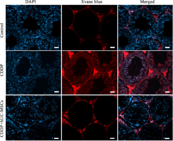
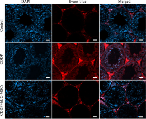
Researchers from the Shandong Stem Cell Engineering Technology Research Center reported that human umbilical cord mesenchymal stem cells (hUC-MSCs) repair cisplatin (CDDP)-induced blood-testis barrier (BTB) damage and restore reproductive function in mice by inhibiting ferroptosis. Following intraperitoneal CDDP injection, mice exhibited abnormal testicular morphology, disorganized seminiferous tubules, reduced sperm count and motility, increased BTB permeability, downregulated BTB-related proteins, and altered ferroptosis markers. Tail vein injection of hUC-MSCs 24 hours after CDDP reversed these effects; the ferroptosis inhibitor Fer-1 mimicked this protection. In vitro fertilization assays showed normalized embryo formation rates in the hUC-MSCs-treated group. In these animal experiments, researchers used ready-to-use 2,2,2-Tribromoethanol (Avertin, AbMole, M2223) as an anesthetic. On day 14, mice were anesthetized with an intraperitoneal dose of 240 mg/kg. Tribromoethanol minimized animal distress, complied with ethical standards, and enabled reliable collection of testicular, epididymal, and sperm samples without tissue damage, which was critical for subsequent histological analysis, protein detection, and sperm quality assessment [6].

Evans blue permeability assay for BTB integrity. Blue fluorescence indicates DAPI-stained nuclei, while red fluorescence corresponds to Evans blue staining

References and Acknowledgments

[1] Q. Chen, M. M. Wells, T. S. Tillman, et al., Structural Basis of Alcohol Inhibition of the Pentameric Ligand-Gated Ion Channel ELIC, Structure (London, England : 1993) 25(1) (2017) 180-187.

[2] A. Luethy, J. D. Boghosian, R. Srikantha, et al., Halogenated Ether, Alcohol, and Alkane Anesthetics Activate TASK-3 Tandem Pore Potassium Channels Likely through a Common Mechanism, Molecular pharmacology 91(6) (2017) 620-629.

[3] Robert E Meyer, Richard E %J Lab animal Fish, A review of tribromoethanol anesthesia for production of genetically engineered mice and rats, 34(10) (2005) 47-52.

[4] William A Hill, Jacquelyn T Tubbs, Christopher L Carter, et al., Repeated administration of tribromoethanol in C57BL/6NHsd mice, 52(2) (2013) 176-179.

[5] Arlene McDowell, Jessica A. Fothergill, Azeem Khan, et al., A cyclodextrin formulation to improve use of the anesthetic tribromoethanol (Avertin®), Journal of Pharmacy and Bioallied Sciences 6(1) (2014).

[6] X. Liu, X. Wang, X. Zhang, et al., Human umbilical cord mesenchymal stem cells ameliorates cisplatin-induced blood-testis barrier dysfunction in mice by mitigating ferroptosis, Stem cell research & therapy 16(1) (2025) 511.

AbMole Mini-Lecture | Treosulfan (Treosulphan): A Highly Efficient DNA Crosslinking Agent and Its Applications in Cancer Research

Treosulfan (Treosulphan, NSC 39069, AbMole, M13635) is an alkylator that undergoes non-enzymatic hydrolysis under physiological conditions to generate active metabolites, exhibiting significant cell proliferation inhibitory effects. It serves as a critical tool in various tumor models and hematopoietic stem cell transplantation studies. The chemical structure of treosulfan features a six-carbon sugar alcohol backbone, in which four hydroxyl groups are methylated, the hydroxyl group at the C2 position is substituted with a sulfhydryl group, and sulfate ester groups are attached to the C1 and C4 positions, respectively. This structural design imparts chemical stability under neutral pH conditions, whereas hydrolysis occurs under weakly acidic environments: the sulfate ester groups are first removed to generate an intermediate monoepoxide derivative (EBDM), which subsequently undergoes spontaneous cyclization to form the electrophilic diepoxide metabolite (DEB). This activation process does not require enzymatic catalysis. As the ultimate active form of treosulfan (NSC 39069), DEB attacks nucleophilic sites (primarily the N7 position of guanine) in DNA molecules, forming intra- and interstrand crosslinks. These crosslinks distort the DNA double helix, impede the normal functions of DNA polymerases and topoisomerases, and ultimately induce S-phase cell cycle arrest and activation of apoptotic pathways. Compared to traditional nitrogen mustard alkylating agents, the diepoxide structure of treosulfan exhibits higher DNA crosslinking efficiency, with IC₅₀ values reaching micromolar or even nanomolar levels in various tumor cell lines.

Treosulfan (Treosulphan, AbMole, M13635) has garnered extensive attention in the field of hematopoietic stem cell transplantation. Treosulfan effectively eliminates host hematopoietic cells while causing minimal damage to non-hematopoietic tissues. For instance, in NOD/SCID mouse models, preconditioning with treosulfan (CAS No.: 299-75-2) (1.5 g/kg/day for 3 days) significantly improves the engraftment success rate of CD34⁺ cells. Beyond hematologic malignancy research, treosulfan (NSC 39069) is also applicable to cell- and animal-level studies of other tumors. For example, treosulfan in combination with fludarabine synergistically kills ovarian cancer cell lines SKOV3 and OVCAR4 [1]. In an Ewing’s sarcoma A673 cell xenograft model, intraperitoneal injection of treosulfan (150 mg/kg, twice weekly) reduces tumor volume by 64% while downregulating the expression of ETS fusion genes [2].

References and Acknowledgments

[1] I. El-Serafi, I. Micallef Nilsson, A. Moter, et al., Impact of fludarabine and treosulfan on ovarian tumor cells and mesothelin chimeric antigen receptor T cells, Cancer immunology, immunotherapy : CII 73(9) (2024) 163.

[2] Sebastian Werner, Arnulfo Mendoza, Ralf A Hilger, et al., Preclinical studies of treosulfan demonstrate potent activity in Ewing’s sarcoma, 62(1) (2008) 19-31.