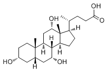
While progress has been made in identifying potential pathways involved in diabetic retinopathy, current treatments remain limited. Our own studies have shown that loss of sympathetic neurotransmission, specifically b-adrenergic receptor signaling, produced retinal changes similar to that observed in diabetic animal models. Likewise, we demonstrated that loss of dopamine beta hydroxylase, b-1-adrenergic receptor signaling, or b-2-adrenergic receptor signaling can all produce a phenotype similar to diabetic retinopathy, in the absence of changes in glucose levels. Based on these findings, we tested whether or not the adrenergic receptor agonist, isoproterenol, could restore normal b-adrenergic receptor signaling in the eye and thus prevent retinal damage associated with diabetes using the streptozotocin-induced diabetes rat model. Results showed that as predicted, isoproterenol treatment prevented retinal damage; however, the treatment also caused unacceptable side effects in the heart. To avoid these cardiovascular changes, we synthesized a novel b-adrenergic receptor agonist, Compound 49b, which selectively prevents both vascular and neuronal changes associated diabetes in the retina but has li le effect on the heart. The discovery of Compound 49b has not only provided an important potential treatment strategy for diabetic retinopathy, it also has provided a useful research tool for further analysis of pathways that alter diabetes-induced changes in retina. Our continuing studies of Compound 49b in the streptozotocininduced diabetic rat model in vivo and in identified retinal cell types in vitro indicate that its likely mechanism of action is through increasing insulin-like growth factor Evodiamine binding protein 3 levels, while decreasing tumor necrosis factor alpha. Our data suggest that the effect of Compound 49b on IGFBP3 does not involve interactions with the insulin-like growth factor, IGF-1, but rather independent actions of IGFBP-3 acting through the IGFBP-3 receptor. We have subsequently demonstrated that Compound 49b regulates IGFBP-3 through DNA-PK to prevent apoptosis of retinal endothelial cells. Additionally, we have shown that IGFBP-3 regulates retinal endothelial cell apoptosis through binding to its receptor. Based upon our data with Compound 49b, we hypothesized that IGFBP 3 is upstream of TNFa, such that maintenance/restoration of IGFBP-3 actions would prevent TNFa��s inhibition of insulin signaling. Our findings of increased TNFa and SOCS3 in retinal endothelial and Mu��ller cells under high glucose conditions agree well with findings in other cell types, including adipocytes and smooth muscle cells, suggesting that these retinal cells may also undergo a form of insulin resistance. In normal insulin signal transduction, autophosphorylation of the insulin receptor on tyrosine 1150/1151 leads to activation of IRS-1 or IRS-2, which phosphorylate Akt, a potent anti-apoptotic factor, thus preventing apoptosis of cells. We have shown that TNFa blocks normal insulin signal transduction in both retinal endothelial and Mu��ller cells. Under high glucose  conditions, TNFa increases phosphorylation of insulin receptor Procyanidin-B2 substrate 1 on serine 307, thus inhibiting the ability of IRS-1 to activate Akt to prevent apoptosis.